Computed tomography – CT

Computed tomograph
Computed tomography - CT 12

Computed tomography vet: A modern diagnostic method for your pet

introduction

Modern veterinary medicine is constantly developing and offers more and more options for diagnosing illnesses in pets early and accurately. One such method is the computer tomography (CT). In this article you will learn how a CT is used by a veterinarian, what advantages this method offers and which clinical pictures can be examined with it.

What is a CT scanner?

A CT scanner is a state-of-the-art medical device that can use X-rays to create three-dimensional images of an animal's interior. This technique allows veterinarians to obtain detailed information about an animal's anatomy and possible diseases without having to perform invasive procedures

When is a CT used at the vet?

A CT scanner is used in various situations to make a precise diagnosis and determine the best possible therapy for your pet. Some of the most common areas of application are:

  • Neurology : Examination of the brain and spinal cord to identify conditions such as tumors, inflammation, or injury.
  • Oncology : Diagnosis of tumors and metastases to determine the exact location, size and extent of cancerous tumors.
  • Orthopedics : Visualization of bones, joints and soft tissues to detect fractures, osteoarthritis or herniated discs.
  • Cardiology : Examination of the heart and blood vessels to diagnose heart defects, heart failure, or blood clots.

Advantages of computed tomography in veterinary medicine

Computed tomography offers several advantages over other imaging procedures such as X-rays or magnetic resonance imaging (MRI):

  1. Speed : The CT scan typically only takes a few minutes, which is particularly beneficial for animals in pain or anxiety.
  2. Precision : The three-dimensional images enable a more precise representation of the anatomy and possible diseases, making diagnosis easier.
  3. Minimally invasive : Since no invasive procedures are required, the animal is not placed under unnecessary stress during the examination.

Process of a CT examination at the vet

Before the CT scan begins, the animal is usually sedated or placed under general anesthesia to ensure that it remains calm during the scan. The animal is then positioned on a special table and pushed into the CT tube. During the examination, the X-ray tube rotates around the animal and takes multiple X-ray images from different angles. The data obtained is then analyzed by a computer and converted into three-dimensional images.

Once the images are created, they are evaluated by a veterinarian or a specialized radiologist. The results of the examination can then be discussed directly with the animal owner and the necessary treatment options can be initiated.

Possible risks and side effects of computer tomography

Although CT is a relatively safe procedure, there are some possible risks and side effects that pet owners should consider:

  1. Radiation exposure : As with any x-ray examination, a certain amount of radiation exposure is also present in computer tomography. However, this is usually very low and poses only minimal risk to the animal.
  2. Anesthesia : Because the CT scan is performed under sedation or general anesthesia, there is some risk of anesthesia-related complications. However, this risk is also low and is minimized by a thorough examination of the animal and its state of health before the examination.
  3. Allergic reactions : In some cases, a CT scan uses a contrast agent to make certain structures more visible. Some animals may experience allergic reactions to the contrast medium. However, these are rare and usually easy to treat.

Cost of a computer tomography at the vet

The cost of a CT scan can vary depending on the veterinary practice, region and the animal's individual circumstances. As a rule, the prices for a CT examination are between 500 and 1,500 euros. In some cases, additional costs may apply for anesthesia, contrast agent or image evaluation. It is therefore advisable to obtain a cost estimate before the examination and find out about any additional costs.

Our computer tomograph iCRo Claris V

State-of-the-art 3D Veterinary Imaging: An Overview of the Claris V CBCT Scanner

Advanced imaging technologies play a crucial role in modern veterinary medicine. The Claris V Cone Beam Computed Tomography (CBCT) scanner is at the forefront of these innovations, providing veterinary professionals with detailed and precise diagnostics. In this article, we will detail the unique features and benefits of the Claris V CBCT scanner.

High-resolution imaging: the key to precise diagnosis

Superior image quality

The Claris V CBCT scanner uses high-resolution detectors and isotropic voxels to produce images of exceptional clarity. These technologies reduce scattering effects and improve image processing, resulting in crisp and accurate 3D images. This high image quality is critical for diagnosing and treating a wide range of veterinary conditions.

Optimized scanning parameters

The CBCT scanner uses optimized scanning parameters to ensure consistent and high quality imaging. These parameters are designed to provide the best possible image resolution with minimal radiation exposure to the patient.

Versatile uses in veterinary medicine

3D CT, X-ray and fluoroscopy

The Claris V CBCT scanner offers triple-play scanning functionality that includes 3D CT, X-ray and fluoroscopy. This versatility allows veterinarians to perform various diagnostic procedures with a single device, significantly improving efficiency and workflow in the practice.

Mobile solution for flexibility

The Claris V Scanner is a mobile solution that can be easily rolled into any room and plugged directly into a wall outlet. This mobility offers veterinary clinics the flexibility to deploy the scanner in different locations without the need for extensive installation.

Benefits for patients and owners

Reduced anesthesia risks

The rapid and precise imaging of the Claris V CBCT scanner can significantly reduce the risks associated with repeated anesthesia. This results in less stress for patients and increases safety during diagnostic and therapeutic procedures.

Stress reduction for patients and owners

The rapid diagnosis and treatment enabled by the use of the Claris V CBCT scanner reduces stress for both animals and their owners. Fast and accurate results help ensure treatment decisions can be made quickly, making the entire treatment process easier.

Areas of application of the Claris V CBCT scanner in veterinary medicine

The Claris V CBCT scanner represents a groundbreaking innovation in veterinary diagnostics. Thanks to its high-resolution imaging and versatile application options, it enables precise diagnosis and treatment of a wide range of animal diseases. In this article we explain the diverse areas of application of this advanced device in veterinary medicine.

Diagnosis and treatment of dental and jaw diseases

Dental root examinations

Dental problems are common in pets. The Claris V CBCT scanner provides detailed 3D images of tooth roots, allowing dentists to detect hidden problems such as infections, fractures or abnormal root canal structures. This enables precise planning and implementation of root canal treatments.

Orthodontic assessment

Misaligned teeth and jaw abnormalities can also be accurately diagnosed. By viewing the entire jaw area in 3D, veterinarians can make a comprehensive assessment and initiate appropriate orthodontic measures.

Examination of head and neck structures

Nasal cavity and sinus examinations

Diseases of the nose and sinuses such as tumors, polyps or chronic inflammation can be examined in detail using the CBCT scanner. The precise 3D representation of these structures enables precise diagnosis and targeted therapeutic interventions.

Ear examinations

Ear diseases, particularly in the middle ear and inner ear, can be difficult to diagnose using traditional methods. The Claris V CBCT scanner allows for a comprehensive examination of these areas, making it easier to diagnose and treat ear infections, tumors or other abnormalities.

Orthopedic applications

Fracture analysis and healing process

The CBCT scanner is an essential tool for analyzing bone fractures. The detailed 3D images help veterinarians assess the severity of the fracture and monitor healing progress. This is particularly important for planning surgical procedures and evaluating postoperative recovery.

Joint examinations

Joint problems such as arthritis, dysplasia or dislocations can also be accurately diagnosed using the CBCT scanner. The high-resolution images enable a detailed examination of the joint structures and support the planning of suitable treatment strategies.

Examination of soft tissues

Tumor and tumor spread analysis

The Claris V CBCT scanner is ideal for examining soft tissue tumors and their spread. Precise imaging helps determine the size, location and depth of infiltration of tumors, which is crucial for planning surgery or radiation therapy.

Examination of organs

Internal organs such as the lungs, liver or kidneys can also be examined in detail using the CBCT scanner. This is particularly useful in diagnosing organ diseases, tumors or other pathological changes.

Emergency and trauma diagnostics

Fast and precise imaging

In emergencies, every minute counts. The Claris V CBCT scanner provides rapid and precise imaging, which is critical for the immediate diagnosis and treatment of trauma. This can save lives and significantly improve the prognosis for seriously injured animals.

Minimizing the duration of anesthesia

Thanks to the rapid imaging processes of the CBCT scanner, the duration of anesthesia for patients can be minimized. This reduces the risk of complications and contributes to the safety and well-being of the animals.

The Claris V CBCT scanner offers an impressive range of applications in veterinary medicine. From detailed diagnostics of dental and jaw disorders to comprehensive examinations of head, neck and soft tissue structures, this advanced device is an essential tool for modern veterinary practices. With its precise and rapid imaging, the CBCT scanner contributes significantly to improving veterinary care and helps veterinarians make informed and effective treatment decisions.

Claris V CBCT scanner diagram
Claris V CBCT scanner
High-resolution imaging
Versatile uses
Benefits for patients and owners
Economical advantages
Isotropic voxels
Optimized scanning parameters
3D CT, X-ray, fluoroscopy
Mobile solution
Reduced anesthesia risks
Stress reduction

What does this mean for your loved one?

✔ faster and less radiation examination

✔ creates real 3D images, which can then be used, for example, to prepare complicated bone fractures using a 3D printer

Sub-disciplines of veterinary medicine such as orthopedics, cardiology, pulmonology and urology, as well as postoperative controls, benefit from his exceptional skills.
The Claris V™ can also do everything any other CT can do. The Claris V™ allows computed tomography imaging of various body regions such as: B. Effectively display the paranasal sinuses and nasal passages, bulla, ear, brain, teeth or spine.

Further advantages of our computer tomograph:

  • Size of isotropic voxels ≥ 90x90x90 µm
  • allows patients to be scanned in their full length
  • Warm-up takes less than 3 minutes
  • enables examinations of patients up to 80 kg
  • was developed for diagnostic, interventional and intraoperative use
  • integrated 2D, MPR and 3D imaging
  • diagnostically advantageous soft tissue contrast
  • Software enables cutting of the 3D images on all levels in excellent quality
  • Export of examination results in standard DICOM format

In addition to our in-house experts, we work closely with internationally trained experts to evaluate the examination results so that you have an optimal examination report in your hands at the end of every CT examination.

Important areas of application for our computer tomograph are:

  • Dental x-rays, e.g. FORL in cats - but also dogs, rabbits and guinea pigs can be examined in more detail using computer tomography
  • Diagnosis of tumors and metastases
  • Setting up bone fractures
  • preoperative planning of fracture treatment
  • Representation of vessels, bile ducts and gastrointestinal sections with appropriate contrast media
  • Placement of probes
  • Observation of dynamic processes
    • Heart movement
    • Swallowing movement (esophagus representation)
  • Integrating implant control during surgery (functions similar to a C-arm)
  • Biopsies
  • Positioning control before the CT examination

In certain areas, our VIMAGO™ is not only equal to a human medical 64-row computer tomograph, but also superior. You can find a comparison here:

Here is an example of a cat dental CT taken with our in-house computer tomograph:

Cat head CT
Cat head CT

And here is the corresponding 3D printout from our in-house 3D printer:
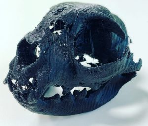
3D printout CT cat skull
Computed tomography - CT 15

Advantages of the Claris V CBCT scanner compared to classic 16-row computer tomographs in veterinary medicine

Computed tomography is an advanced and precise method for diagnosing diseases in pets. It allows veterinarians to obtain detailed information about an animal's anatomy and possible diseases without having to perform invasive procedures. The CT scan is quick, minimally invasive and generally safe. However, pet owners should inform themselves about possible risks, side effects and costs before deciding to undergo a CT scan.

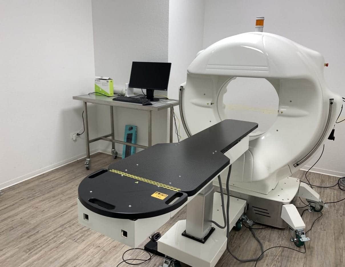
Computed tomography (CT) has established itself as a valuable diagnostic tool in veterinary medicine. While traditional 16-slice CT scanners are widely used, the Claris V CBCT (Cone Beam Computed Tomography) scanner offers numerous advantages that make it a superior choice. In this article we explain the differences and the specific advantages of the Claris V CBCT scanner compared to classic 16-row CT scanners.

Technology and Imaging

Different radiation techniques

A key difference between CBCT and traditional 16-slice CT scanners is the way the images are generated. While 16-row CT scanners use multiple rows of detectors to create slice-by-slice images, the CBCT scanner uses a cone-shaped X-ray beam and a single detector to capture volumetric 3D images in a single scan.

Higher resolution and image quality

The Claris V CBCT scanner offers superior image quality with higher resolution. The isotropic voxels and advanced image processing algorithms ensure more detailed and precise 3D images. This is particularly beneficial for examining small structures and fine anatomical details that may not be as clearly visible with a 16-row CT.

Scan efficiency and patient comfort

Faster scanning times

The CBCT scanner requires less time for a complete scan due to its volumetric imaging technology. This significantly reduces the total time an animal must remain calm or be sedated. Faster scanning times mean less stress for the animal and more efficient workflows in the veterinary practice.

Lower radiation exposure

Another significant advantage of the CBCT scanner is the lower radiation exposure compared to conventional 16-row CT scanners. Because the CBCT scanner uses a cone-shaped beam and requires fewer frames, radiation exposure to the animal is minimized. This is particularly important for repeated examinations or for sensitive patients.

Mobility and flexibility

Compact and mobile design

The Claris V CBCT scanner is more compact and portable than many traditional 16-row CT scanners. It can be easily moved to different rooms and plugged directly into a regular power outlet. This mobility offers veterinary practices and clinics greater flexibility in using the scanner and saves valuable space.

Easy integration and plug and play

Thanks to its plug-and-play functionality, the CBCT scanner can be quickly and easily integrated into everyday practice. Unlike the often more complex installation requirements of a 16-slice CT scanner, the Claris V can be quickly deployed, further increasing practice efficiency.

Specific applications and versatility

Extensive diagnostic options

While traditional 16-slice CT scanners are primarily used for general imaging purposes, the Claris V CBCT scanner offers a wider range of specific applications. This includes detailed examinations of the tooth and jaw structures, the nasal cavities and sinuses, as well as the complex head and neck anatomy.

Versatile imaging modes

The Claris V CBCT scanner supports X-ray and fluoroscopy modes in addition to 3D imaging. This versatility makes it an extremely valuable tool for veterinarians who want to perform various diagnostic procedures with a single device.

The Claris V CBCT scanner offers numerous advantages over traditional 16-slice CT scanners, making it a superior choice for veterinary diagnostics. With its higher image quality, lower radiation exposure, faster scan times and greater flexibility, the CBCT scanner represents a significant improvement. These advantages help increase diagnostic accuracy, improve patient comfort and increase practice efficiency. Veterinarians who invest in a Claris V CBCT scanner can be confident that they are providing their patients with the best possible diagnostic care.

Catchment area for our CT within a radius of 30 km around Karlsruhe

Our state-of-the-art Claris V CBCT scanner is not only available to veterinarians in Karlsruhe, but also serves a large catchment area within a 30-kilometer radius. The towns and communities in this catchment area include Ettlingen, Rheinstetten, Stutensee, Bruchsal, Durlach, Weingarten (Baden), Pfinztal, Germersheim, Waghäusel, Walzbachtal, Malsch, Durmersheim, Bad Schönborn, Philippsburg, Graben-Neudorf, Eggenstein-Leopoldshafen, Forst , Kronau, Linkenheim-Hochstetten, Östringen, Rastatt, Sulzfeld, Karlsbad, Bretten and Wörth am Rhein.

Pet owners from these locations can easily access our advanced imaging technology, enabling precise diagnosis and treatment of their pets. Our central location in Karlsruhe ensures that high-quality veterinary care is also accessible to the surrounding communities and cities. We look forward to supporting and promoting the health and wellbeing of animals across the region.

Conclusion

The computer tomograph: An indispensable tool in modern veterinary medicine

A computer tomograph is an indispensable instrument in modern veterinary medicine. The computer tomograph enables detailed and precise diagnoses that are often not possible with conventional methods. Thanks to the advanced technology of a CT scanner, veterinarians can gain deeper insight into their patients' health. Our computer tomograph in Karlsruhe also offers lower radiation exposure for the animals.

A computer tomograph not only improves the accuracy of diagnosis, but also the efficiency of treatments. With a computer tomograph, complex medical conditions can be identified quickly and reliably. The mobility of our computer tomograph also makes it easier to access in different practices. By using a computer tomograph, repeated anesthesia can be avoided.

Every computer tomograph in our practice is regularly maintained to ensure the highest quality. The computer tomograph is therefore an essential part of veterinary care. Overall, it shows that computer tomography plays a crucial role in improving the diagnosis and treatment of animals.

Scroll up