Cauda Equina Syndrome: Comprehensive information on diagnosis and treatment
- Cauda Equina Syndrome: Comprehensive information on diagnosis and treatment
- Overview of Cauda Equina Syndrome
- Explanation of Cauda Equina Syndrome
- Symptoms of Cauda Equina Syndrome
- Diagnostic methods
- Treatment options
- Rehabilitation phase
- Comprehensive care during and after treatment
- Frequently asked questions about Cauda Equina Syndrome - FAQs:
- Summary
Overview of Cauda Equina Syndrome
Cauda equina syndrome is a progressive neurological disease characterized by narrowing of the nerve roots in the lumbar spine and sacrum. This disease mainly affects dogs of large, sporting breeds from middle age onwards. Symptoms are varied and can range from pain to lameness to paralysis and incontinence.
Explanation of Cauda Equina Syndrome
Cauda Equina Syndrome occurs when the nerve roots in the lumbar-sacral region are compressed. This condition is particularly common in larger sporting breed dogs that have reached middle age.

Symptoms of Cauda Equina Syndrome
In many cases, Cauda Equina Syndrome can go undetected for a long time because dogs often suffer patiently despite increasing pain. Symptoms that may occur include:
- Alternating lameness in one or both hind limbs
- Stiff gait
- Chewing on the tail or hind legs
- Difficulty standing up on the hind legs and maintaining a stretched posture
- Vocalizations with moans or whimpers or with sudden screams or howls
- Paralysis of the hind limbs, sphincters and bladder (fecal and urinary incontinence)
Diagnostic methods
Neurological examination
The neurological examination includes the assessment of the movement sequence at walk, trot and canter as well as special examinations for pain and neurological dysfunction in order to localize the location of the nerve block more precisely.
Imaging procedures
- Direct digital X-ray: Provides better visualization of bones and soft tissues, higher contrast resolution, and the ability to enlarge images and change brightness.
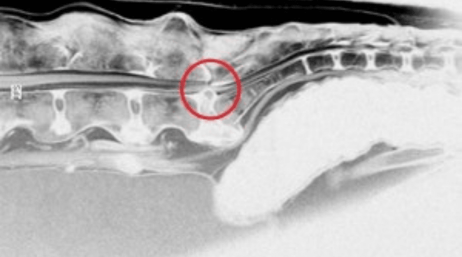
- Myelography or epidurography: Contrast X-ray examinations of the spinal canal to detect changes in the spinal canal and vertebrae and if disc herniations are suspected.
- Computed tomography (CT): Allows a precise representation of the anatomical structures and the creation of three-dimensional images of the changes. CT is particularly helpful in planning any surgical procedure that may be necessary.
Treatment options
Conservative therapy
For patients in the first stage of the disease and with only minor pain, medical treatment with anti-inflammatory, pain-relieving medications and rest for the patient can be tried.
Surgical therapy
Surgical therapy is recommended for neurological deficits and pain that do not respond to painkillers or recur. A dorsal laminectomy is performed to relieve pressure on the trapped nerve roots.
Risks of cauda equina surgery / dorsal laminectomy
As with any surgery, there may be risks and complications associated with a dorsal laminectomy to treat cauda equina syndrome in dogs. Here are some of the possible risks:
- Anesthesia Risks: As with any surgical procedure, there is a risk of complications related to anesthesia, including allergic reactions and breathing problems.
- Bleeding: Bleeding may occur during surgery and can be controlled in most cases. However, in rare cases, severe bleeding may occur and may require a transfusion.
- Infection: A postoperative infection can occur in the area of the surgical site. Usually this can be treated with antibiotics, but in some cases additional surgery may be required.
- Nerve damage: There is a risk of nerve damage with a dorsal laminectomy. This can lead to persistent pain, weakness or numbness in the hind legs, as well as impaired bladder and bowel function.
- Cauda Equina Syndrome Recurrence: In some cases, cauda equina syndrome may recur after surgery if the underlying causes have not been fully resolved.
- Spinal instability: Removal of portions of the spinal canal can lead to spinal instability. In some cases, this may require the need for further surgery to stabilize the spine.
- Scarring: As with any surgery, scarring can occur with a dorsal laminectomy. This can lead to persistent pain or restricted movement.
It is important to note that the risk of complications depends on several factors, including the dog's health, the severity of the cauda equina syndrome, and the experience of the surgeon. Careful preoperative diagnostics and choosing an experienced surgeon can help minimize the risk of complications.
Rehabilitation phase
Immobilizing the patient for 6 weeks is crucial for rehabilitation. Strenuous activities must be avoided during this time.
During this time, various measures are taken to help the affected animals heal and improve their quality of life. Some of the most important aspects of the rehabilitation period are:
- Pain Management: Pain relief is a key factor in the treatment of dogs with Cauda Equina Syndrome. The veterinarian will usually prescribe painkillers to help relieve the animal's discomfort and allow it to move better.
- Physiotherapy: Physiotherapeutic measures are an important complement to medical treatment and can help to improve the mobility and muscle strength of affected dogs. This may include core strengthening exercises, massage, and passive limb movements.
- Weight management: Excess weight can increase stress on the spine and delay recovery. Therefore, it is important that the dog maintains a healthy weight. A balanced diet and regular, appropriate exercise are crucial here.
- Adjusting the home environment: The dog's environment should be designed to support his recovery. This includes avoiding slippery floors, offering soft lying surfaces and providing ramps to avoid climbing stairs.
- Regular check-ups: It is important that the dog is regularly examined by the veterinarian during the rehabilitation period. This allows progress to be monitored and treatment adjusted if necessary.
- Patience and Support: Recovering from Cauda Equina Syndrome can take time and requires patience and support from the pet owner. A loving and understanding approach helps the dog recover more quickly and improves its well-being.
Overall, the rehabilitation phase is a crucial factor for the successful treatment of cauda equina syndrome in dogs. Close cooperation between veterinarian and animal owner as well as individually tailored therapy are of central importance in order to improve the quality of life of the affected animals and minimize possible neurological damage.
Interdisciplinary approaches to treatment
The collaboration between different specialist areas plays an important role in the treatment of Cauda Equina Syndrome. These include neurosurgeons, orthopedists, radiologists, physiotherapists, urologists and neuropsychologists. This interdisciplinarity enables holistic care for the patient, which addresses not only the physical but also the psychosocial aspects of the disease.
The involvement of urology is particularly relevant, as cauda equina syndrome is often associated with urological problems such as urinary incontinence or sexual dysfunction. Regular urological evaluation and monitoring are therefore essential.
Neuropsychological care is also key, as patients with cauda equina syndrome often experience significant anxiety and stress. Effective neuropsychological intervention can help mitigate these negative emotional reactions and improve patients' quality of life.
Comprehensive care during and after treatment
It is important that comprehensive care is provided both during treatment and subsequent rehabilitation. This includes close cooperation between the veterinarian and the pet owner in order to optimally structure the recovery process. Physiotherapy measures can also be a valuable adjunct to promote healing and restoration of nerve function.
Early detection and prevention of Cauda Equina Syndrome
In order to minimize the risk of Cauda Equina Syndrome, it is advantageous to recognize the disease early and take appropriate preventative measures. veterinarian immediately . A regular veterinary examination, especially for dogs belonging to the affected breeds, can help to identify possible problems at an early stage and, if necessary, to take countermeasures.
Frequently asked questions about Cauda Equina Syndrome - FAQs:
What are the most common symptoms of Cauda Equina Syndrome in dogs?
The most common symptoms of cauda equina syndrome in dogs include lower back pain, difficulty standing up or lying down, uncoordinated movements, weakness or lameness in the hind legs, loss of muscle mass in the hindquarters, incontinence, and difficulty defecating.
How is Cauda Equina Syndrome diagnosed in dogs?
Diagnosis of cauda equina syndrome in dogs is usually made through a thorough clinical examination, which may include neurological testing, x-rays, computed tomography (CT), or magnetic resonance imaging (MRI). These examinations allow the veterinarian to identify possible causes of the symptoms and initiate appropriate treatment.
How can I help my dog recover from Cauda Equina Syndrome?
To help your dog recover from Cauda Equina Syndrome, you should carefully follow your veterinarian's instructions regarding pain management, physical therapy, and weight management. In addition, it is important to adapt the home environment accordingly to make everyday life easier for the dog. Regular check-ups with the vet are also important to monitor recovery progress and adjust treatment if necessary. Finally, patience, love and support will also help your dog recover faster and improve his well-being.
This diagram shows the process from recognizing the signs of Cauda Equina Syndrome to restoring the affected dog's quality of life. Successful treatment and rehabilitation are crucial to maintaining the dogs' quality of life and minimizing neurological damage.
Summary
Cauda Equina Syndrome is a painful and progressive disease that occurs primarily in large breeds of dogs. Diagnosis and treatment requires an experienced veterinarian and may involve different therapeutic approaches depending on the severity of the disease. Early detection and treatment are essential to maintain the quality of life of affected dogs and minimize possible neurological damage.
Current research on Cauda Equina Syndrome in dogs
In recent years, research into cauda equina syndrome in dogs has made significant progress. Some of the recent developments and findings include:
- Improved imaging techniques: New technologies such as magnetic resonance imaging (MRI) and computed tomography (CT) allow for more accurate diagnosis and localization of affected nerve roots, making treatment more effective.
- Minimally Invasive Surgical Procedures: Advances in surgery have led to the development of minimally invasive techniques that are less traumatic for the dog and allow for a quicker recovery.
- Biomarker research: Scientists are looking for biomarkers that could be helpful in the early detection of cauda equina syndrome. This could help detect disease progression in a timely manner and initiate appropriate treatments.
- Stem cell therapy: Some studies are investigating the use of stem cells to treat nerve damage caused by cauda equina syndrome. Although this is still in the experimental phase, there is hope that stem cell therapy could be an effective treatment method in the future.
- Physiotherapy and Rehabilitation: New insights into the importance of physiotherapy and rehabilitation in the recovery of dogs with Cauda Equina Syndrome have led to the development of individual rehabilitation plans to support the healing process and improve the quality of life of the affected animals.
Research into cauda equina syndrome in dogs continues to evolve, and further advances in diagnosis, treatment, and rehabilitation are expected to occur in the coming years.
outlook
There are still many open questions and challenges in the area of cauda equina syndrome in dogs that researchers need to further investigate in the future:
- Genetics: Research into the genetic causes and risk factors for cauda equina syndrome in dogs is an important step toward better understanding the disease and potentially developing preventive measures.
- Prevention: Because some dog breeds are more susceptible to Cauda Equina Syndrome, it is important to develop strategies to prevent the disease in these breeds. This may include identifying risk factors, implementing breeding programs to reduce incidence, and educating dog owners.
- Long-term studies: Further long-term studies are needed to better understand the long-term effects and success of different treatment approaches in dogs with cauda equina syndrome. This will help determine the best treatment options for affected animals.
- Interdisciplinary Collaboration: Close collaboration between veterinarians, surgeons, physical therapists and other professionals is critical to identifying and implementing the best treatment options for dogs with Cauda Equina Syndrome.
- Education and Awareness: Raising public and dog owner awareness of Cauda Equina Syndrome and its potential signs and symptoms is critical to enable early diagnosis and treatment.
Overall, research in the area of cauda equina syndrome in dogs has already made significant progress, but there are still many open questions and challenges that future research projects must address. By continuing to develop diagnostic and treatment methods, as well as improving rehabilitation and prevention strategies, we can hopefully continue to improve the quality of life of dogs affected by this condition.
Literature on the Cauda Equina Syndrome
Here are some recommended literature sources on the subject of Cauda Equina Syndrome:
- Gardner, A., Gardner, E., & Morley, T. (2011). Cauda equina syndrome: a review of the current clinical and medico-legal position. European Spine Journal , 20(5), 690-697.
- Korse, NS, Pijpers, JA, van Zwet, E., Elzevier, HW, & Vleggeert-Lankamp, CL (2017). Voiding disorders as an indicator of cauda equina syndrome: a systematic review. Neurology and Urodynamics , 36(3), 615-623.
- Fraser, S., Roberts, L., & Murphy, E. (2009). Cauda equina syndrome: a literature review of current clinical and medical positions. Emergency Medicine Journal , 26(12), 833-836.
- Podnar, S. (2007). Epidemiology of cauda equina and conus medullaris lesions. Muscles & Nerves , 35(4), 529-531.
- Ahn, UM, Ahn, NU, Buchowski, JM, Garrett, ES, Sieber, AN, & Kostuik, JP (2000). Cauda equina syndrome secondary to lumbar disc herniation: a meta-analysis of surgical outcomes. Spine , 25(12), 1515-1522.
Please note that you will need access to academic journals or academic databases to view some of these sources. You could also use the services of your local academic library to access these publications. These references are only a small selection of the resources available on the topic of Cauda Equina Syndrome, so it is recommended that you conduct further research to get a full picture of this complex topic.
